-
×
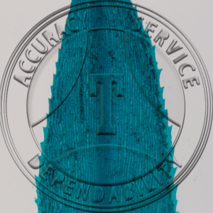
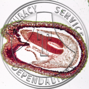
 Tilia americana Flower Bud Median LS Prepared Microscope Slide 17-381A-1
1 × $11.00
Tilia americana Flower Bud Median LS Prepared Microscope Slide 17-381A-1
1 × $11.00 -
×
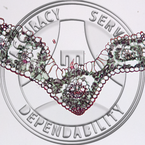
 Capsella bursa-pastoris Embryo Early Cotyledons Non Median Prepared Microscope Slide 20-2BF
1 × $5.25
Capsella bursa-pastoris Embryo Early Cotyledons Non Median Prepared Microscope Slide 20-2BF
1 × $5.25 -
×
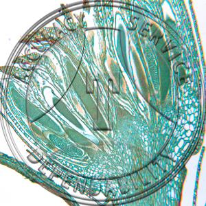
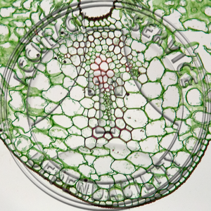 Lilium Leaf Prepared Microscope Slide 16-3A
1 × $5.50
Lilium Leaf Prepared Microscope Slide 16-3A
1 × $5.50 -
×
 Obelia Gonangium Prepared Microscope Slide ZC2-1
1 × $9.60
Obelia Gonangium Prepared Microscope Slide ZC2-1
1 × $9.60 -
×
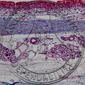 Pseudostratified Ciliated Columnar Epithelium Trachea Prepared Microscope Slide HA3-32
1 × $7.25
Pseudostratified Ciliated Columnar Epithelium Trachea Prepared Microscope Slide HA3-32
1 × $7.25 -
×
 Polytrichum Mature Capsule CS Prepared Microscope Slide 6-2DD
1 × $5.50
Polytrichum Mature Capsule CS Prepared Microscope Slide 6-2DD
1 × $5.50 -
×
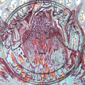
 Iris Leaf Prepared Microscope Slide 16-3C
1 × $6.00
Iris Leaf Prepared Microscope Slide 16-3C
1 × $6.00 -
×
 Polytrichum Young Sporphyte CS Prepared Microscope Slide 6-2F
2 × $6.25
Polytrichum Young Sporphyte CS Prepared Microscope Slide 6-2F
2 × $6.25 -
×
 Crayfish Hemotoxylin Testis Prepared Microscope Slide ZK1-419
1 × $9.25
Crayfish Hemotoxylin Testis Prepared Microscope Slide ZK1-419
1 × $9.25 -
×
 Lilium Leaf Epidermis Prepared Microscope Slide 16-3B
1 × $5.50
Lilium Leaf Epidermis Prepared Microscope Slide 16-3B
1 × $5.50 -
×
Pennaria Gonophores Prepared Microscope Slide ZC2-31
1 × $10.20
-
×
 Elodea Hydrophytic Leaf Prepared Microscope Slide 16-2A
1 × $5.50
Elodea Hydrophytic Leaf Prepared Microscope Slide 16-2A
1 × $5.50 -
×
 Iris Leaf Epidermis Prepared Microscope Slide 16-3D
1 × $6.00
Iris Leaf Epidermis Prepared Microscope Slide 16-3D
1 × $6.00
Subtotal: $99.05