-
×
 Grass Leaf Prepared Microscope Slide 16-4SP
1 × $6.00
Grass Leaf Prepared Microscope Slide 16-4SP
1 × $6.00 -
×
 Malphigian Tubules Prepared Microscope Slide ZK3-16
1 × $7.00
Malphigian Tubules Prepared Microscope Slide ZK3-16
1 × $7.00 -
×
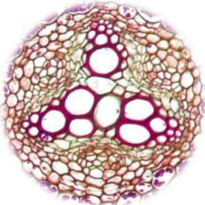
 Pogonatum Stem CS Prepared Microscope Slide A-210A-1
1 × $8.00
Pogonatum Stem CS Prepared Microscope Slide A-210A-1
1 × $8.00 -
×
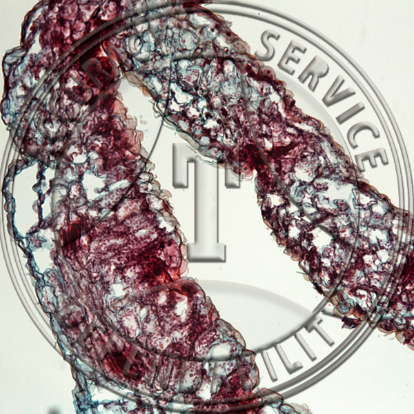
Pennaria Gonophores Prepared Microscope Slide ZC2-31
1 × $10.20
-
×
 Human Louse Male WM Prepared Microscope Slide ZK4-11
1 × $7.00
Human Louse Male WM Prepared Microscope Slide ZK4-11
1 × $7.00 -
×
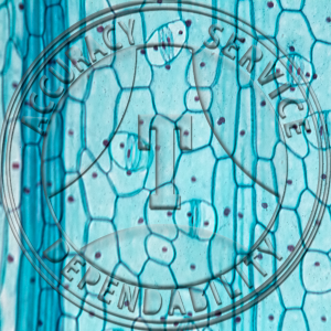
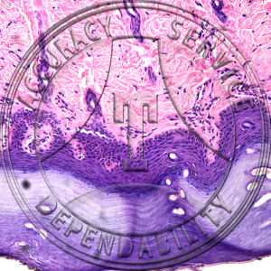 Squamous Epithelium Cornified Prepared Microscope Slide HA4-23
1 × $6.90
Squamous Epithelium Cornified Prepared Microscope Slide HA4-23
1 × $6.90 -
×
Squash Bug Wings Prepared Microscope Slide ZK5-26
1 × $6.00
-
×
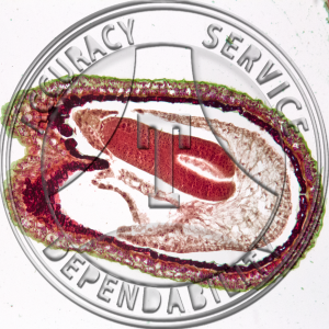
 Mussel Mantel Prepared Microscope Slide ZI1-13
1 × $5.75
Mussel Mantel Prepared Microscope Slide ZI1-13
1 × $5.75 -
×
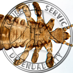
 Bed Bug Prepared Microscope Slide ZK5-1
1 × $7.75
Bed Bug Prepared Microscope Slide ZK5-1
1 × $7.75 -
×
 Triticum aestivum Leaf Prepared Microscope Slide 16-5
1 × $5.50
Triticum aestivum Leaf Prepared Microscope Slide 16-5
1 × $5.50 -
×
 Yucca Leaf Fiber Prepared Microscope Slide 16-8
1 × $5.50
Yucca Leaf Fiber Prepared Microscope Slide 16-8
1 × $5.50
Subtotal: $75.60