-
×
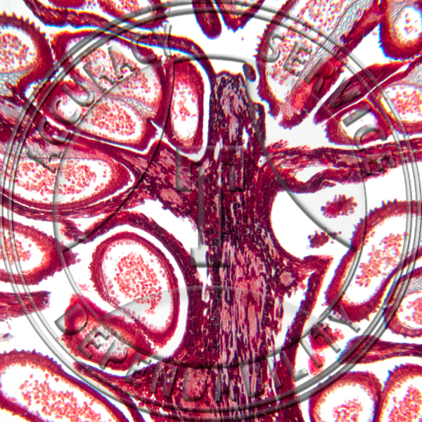
 Polygala polygama Cleistogamous Flower Median LS Prepared Microscope Slide 17-357-2
1 × $13.25
Polygala polygama Cleistogamous Flower Median LS Prepared Microscope Slide 17-357-2
1 × $13.25 -
×
 Lilium Leaf Prepared Microscope Slide 16-3A
2 × $5.50
Lilium Leaf Prepared Microscope Slide 16-3A
2 × $5.50 -
×
Termite Nymph Prepared Microscope Slide ZK3-634
2 × $5.75
-
×
 Elodea Hydrophytic Leaf Prepared Microscope Slide 16-2A
1 × $5.50
Elodea Hydrophytic Leaf Prepared Microscope Slide 16-2A
1 × $5.50 -
×
 Yucca Leaf Fiber Prepared Microscope Slide 16-8
1 × $5.50
Yucca Leaf Fiber Prepared Microscope Slide 16-8
1 × $5.50 -
×
 Pisum sativum Tendril Section Prepared Microscope Slide 15-355-2
1 × $8.20
Pisum sativum Tendril Section Prepared Microscope Slide 15-355-2
1 × $8.20 -
×
 Capsella bursa-pastoris Embryo Early Cotyledons Non Median Prepared Microscope Slide 20-2BF
2 × $5.25
Capsella bursa-pastoris Embryo Early Cotyledons Non Median Prepared Microscope Slide 20-2BF
2 × $5.25 -
×
 Grass Leaf Prepared Microscope Slide 16-4SP
1 × $6.00
Grass Leaf Prepared Microscope Slide 16-4SP
1 × $6.00 -
×
 Phormium Leaf Fiber Prepared Microscope Slide 16-9
1 × $6.00
Phormium Leaf Fiber Prepared Microscope Slide 16-9
1 × $6.00 -
×
 Capsella bursa-pastoris Embryo Bending Cotyledons Median Except Suspensor Prepared Microscope Slide 20-2CC
1 × $23.00
Capsella bursa-pastoris Embryo Bending Cotyledons Median Except Suspensor Prepared Microscope Slide 20-2CC
1 × $23.00 -
×
 Squamous Epithelium Frog Skin Section Prepared Microscope Slide HA4-21
1 × $6.00
Squamous Epithelium Frog Skin Section Prepared Microscope Slide HA4-21
1 × $6.00 -
×
 Iris Leaf Epidermis Prepared Microscope Slide 16-3D
1 × $6.00
Iris Leaf Epidermis Prepared Microscope Slide 16-3D
1 × $6.00 -
×
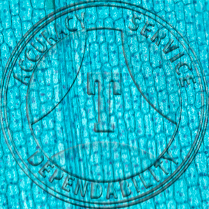
Cat-tail Leaf Prepared Microscope Slide 16-270A
1 × $7.00
-
×
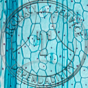
 Lilium Leaf Epidermis Prepared Microscope Slide 16-3B
1 × $5.50
Lilium Leaf Epidermis Prepared Microscope Slide 16-3B
1 × $5.50 -
×
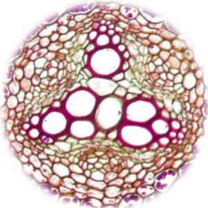
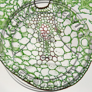
Pogonatum Stem CS Prepared Microscope Slide A-210A-1
1 × $8.00
-
×
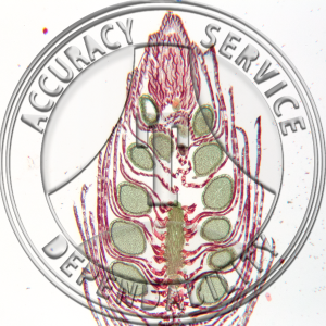
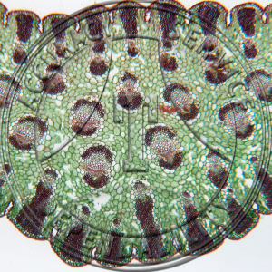
 Tsuga canadensis Male Strobilus LS Prepared Microscope Slide A-251-4
1 × $8.20
Tsuga canadensis Male Strobilus LS Prepared Microscope Slide A-251-4
1 × $8.20 -
×
 Goblet Cells Prepared Microscope Slide HA3-33
1 × $7.25
Goblet Cells Prepared Microscope Slide HA3-33
1 × $7.25 -
×
 Sphagum Mature Sporophyte Median LS Prepared Microscope Slide 6-6A
1 × $14.75
Sphagum Mature Sporophyte Median LS Prepared Microscope Slide 6-6A
1 × $14.75
Subtotal: $163.15