-
×
 Tradescantia Leaf Prepared Microscope Slide 16-4B
1 × $6.00
Tradescantia Leaf Prepared Microscope Slide 16-4B
1 × $6.00 -
×
 Folded & Open Grass Leaf Prepared Microscope Slide 16-4
2 × $9.25
Folded & Open Grass Leaf Prepared Microscope Slide 16-4
2 × $9.25 -
×
 Grass Leaf Prepared Microscope Slide 16-4SP
1 × $6.00
Grass Leaf Prepared Microscope Slide 16-4SP
1 × $6.00 -
×
 Lilium Leaf Prepared Microscope Slide 16-3A
2 × $5.50
Lilium Leaf Prepared Microscope Slide 16-3A
2 × $5.50 -
×
 Bacillus anthracis Mouse Section Prepared Microscope Slide 4-62 sp
1 × $10.00
Bacillus anthracis Mouse Section Prepared Microscope Slide 4-62 sp
1 × $10.00 -
×
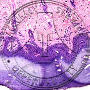 Squamous Epithelium Cornified Prepared Microscope Slide HA4-23
1 × $6.90
Squamous Epithelium Cornified Prepared Microscope Slide HA4-23
1 × $6.90 -
×
Salmonella typhosa Prepared Microscope Slide 4-82c
1 × $5.50
-
×
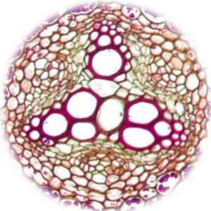
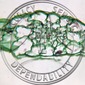 Elodea Hydrophytic Cross Section Prepared Microscope Slide 16-2B
2 × $5.25
Elodea Hydrophytic Cross Section Prepared Microscope Slide 16-2B
2 × $5.25 -
×
Staphylococcus albus Prepared Microscope Slide 4-86a
2 × $5.00
-
×
Pennaria Gonophores Prepared Microscope Slide ZC2-31
1 × $10.20
-
×
Alcaligenes Prepared Microscope Slide 4-60*
1 × $5.50
-
×
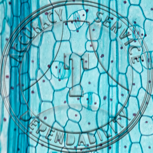 Tradescantia Leaf Stomata Prepared Microscope Slide 16-4A
1 × $5.25
Tradescantia Leaf Stomata Prepared Microscope Slide 16-4A
1 × $5.25 -
×
Polygala polygama Cleistogamous Flower Median LS Prepared Microscope Slide 17-357-2
1 × $13.25
-
×
 Polytrichum Young Sporphyte CS Prepared Microscope Slide 6-2F
1 × $6.25
Polytrichum Young Sporphyte CS Prepared Microscope Slide 6-2F
1 × $6.25 -
×
Clostridium sporogenes Prepared Microscope Slide 4-64aaa
1 × $5.50
-
×
 Polytrichum Young Capsule CS Prepared Microscope Slide 6-2D
1 × $6.00
Polytrichum Young Capsule CS Prepared Microscope Slide 6-2D
1 × $6.00 -
×
 Cuboidal Epithelium Human Kidney Prepared Microscope Slide HA2-11
1 × $9.60
Cuboidal Epithelium Human Kidney Prepared Microscope Slide HA2-11
1 × $9.60 -
×
Modern Cytology Slide Set SET NO.21M MODERN CYTOLOGY; 25 SLIDES
1 × $240.00
-
×
 Slug Slide Prepared Microscope Slide ZI4-2
1 × $5.50
Slug Slide Prepared Microscope Slide ZI4-2
1 × $5.50
Subtotal: $391.45

;-cs")