-
×
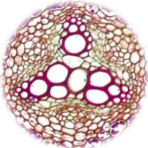
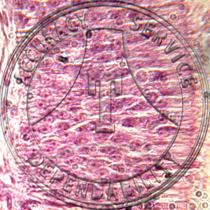
 Epithelium 3 Types Prepared Microscope Slide HA6-11
1 × $9.60
Epithelium 3 Types Prepared Microscope Slide HA6-11
1 × $9.60 -
×
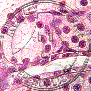
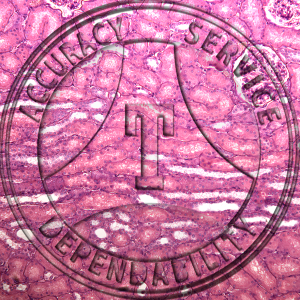 Squamous Epithelium Human Kidney Prepared Microscope Slide HA1-31
1 × $9.60
Squamous Epithelium Human Kidney Prepared Microscope Slide HA1-31
1 × $9.60 -
×
 Psuedostratified Ciliated Columnar Epithelium Nasal Mucosa Prepared Microscope Slide HA3-321
1 × $8.10
Psuedostratified Ciliated Columnar Epithelium Nasal Mucosa Prepared Microscope Slide HA3-321
1 × $8.10 -
×
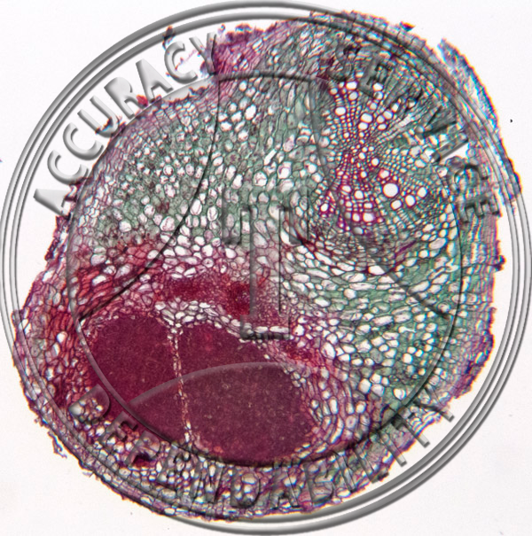
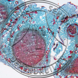
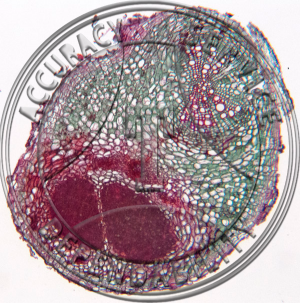 Glycine max Root with Attached Nodule Prepared Microscope Slide 3-94C
1 × $8.25
Glycine max Root with Attached Nodule Prepared Microscope Slide 3-94C
1 × $8.25
Subtotal: $35.55