-
×
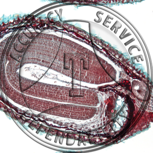
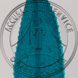 Pogonatum Leaf WM Prepared Microscope Slide A-210A-3
1 × $8.00
Pogonatum Leaf WM Prepared Microscope Slide A-210A-3
1 × $8.00 -
×
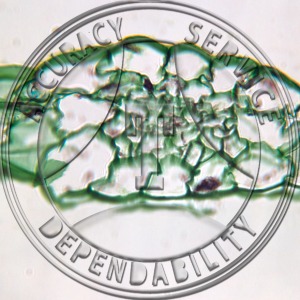
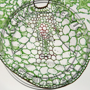 Lilium Leaf Prepared Microscope Slide 16-3A
1 × $5.50
Lilium Leaf Prepared Microscope Slide 16-3A
1 × $5.50 -
×
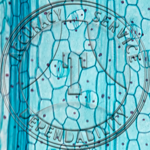
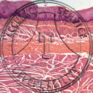 Squamous Epithelium Human Esophagus Prepared Microscope Slide HA4-221
1 × $9.60
Squamous Epithelium Human Esophagus Prepared Microscope Slide HA4-221
1 × $9.60 -
×
 Diplococcus Prepared Microscope Slide 4-68*
1 × $5.25
Diplococcus Prepared Microscope Slide 4-68*
1 × $5.25 -
×
 Sphagum Prothallium WM Prepared Microscope Slide 6-6F
1 × $7.25
Sphagum Prothallium WM Prepared Microscope Slide 6-6F
1 × $7.25 -
×
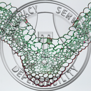
 Folded & Open Grass Leaf Prepared Microscope Slide 16-4
1 × $9.25
Folded & Open Grass Leaf Prepared Microscope Slide 16-4
1 × $9.25
Subtotal: $44.85